

Mammalian ovarian operating manual decoded
The general outline of how immature egg cells are assisted by specific ovarian helper cells is well understood. However, the genetics of the process has been somewhat obscure. Now, new research reveals in startling detail the genetic script that mammalian immature eggs follow as they develop.
Research conducted at the Carnegie Institution of Science, Baltimore, US has provided insights into the maturation of somatic and germ cells of the developing mouse ovary that assemble into ovarian follicles, by determining cellular gene expression and tracing the lineage relationships of the cells1.
Development of ovarian somatic cells
The investigators studied the expression differences between pregranulosa cells of wave 1 follicles (those that function at puberty) and those of wave 2 follicles (those that sustain fertility). This research casts light on the development of ovarian somatic cells and provides a new basis to study the development, physiology and evolutionary conservation of mammalian ovarian follicle formation.
Before birth, germ cells assemble a finite number of cell clusters (i.e. follicles) in the female ovaries. These consist of an immature oocyte together with helper cells, which assist the oocyte through the maturation process. In the final stage during ovulation, the matured oocyte emerges from the follicle.
Understanding the development of follicles
Professor Alan Spradling, Co-investigator explained, “Follicles are slowly used up during a female’s reproductive lifespan and menopause ensues when they run out. Understanding what it takes for follicles to form and develop successfully, helps us learn how damaged genes or adverse environmental factors, including a poor diet, can interfere with fertility.”
He continued, “By documenting the follicle’s genetic operating manual, problems in egg development that might lead to birth defects, as a result of mutations or due to bad nutrition can be better understood and reduced.”
Professor Spradling, and his colleague at the Institution of Science, Dr Wanbao Niu, sequenced 52,500 mouse ovarian cells at 7 stages of follicle development. This allowed them to determine the relative expression of thousands of genes and to characterise their roles. The study illuminated the two distinct types2 of follicles produced by mammals (including humans) and the researchers were able to identify many differences in gene activity between them.
Study looked at distinct types of follicle
- Wave 1 follicles: these are present in the ovary before puberty. In mice, they generate the first fertile eggs. However, their function in humans is poorly understood, but it is thought that they may produce useful hormones
- Wave 2 follicles: these are stored in a resting state, but small groups are activated to mature during a female’s hormonal cycle, ending in ovulation.
Model of epithelial progenitor cells underlying Wave 1 and Wave 2 follicle production
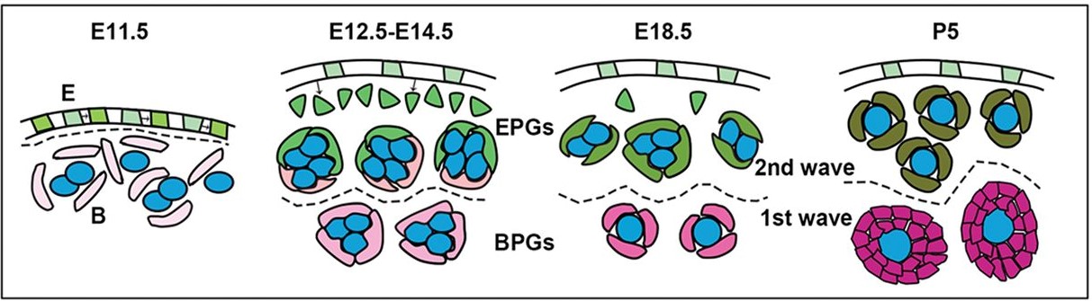
E = embryonic day of study
EPG = epithelial pregranulosa cells
BPG = bipotential pregranulosa cells
BPG cells derive directly from bipotential precursors and are associated with cysts throughout the ovary by E12.5. A second PG group, EPG cells, arise in the ovarian surface epithelium and ingress cortically by E12.5 or earlier.
Professor Spradling said, “We hope our work will serve as a genetic resource for all researchers who study reproduction and fertility.”
References
- Wanbao Niu & Allan C. Spradling. 2020. Two distinct pathways of pregranulosa cell differentiation support follicle formation in the mouse ovary. Proceedings of the National Academy of Sciences. Aug 2020, 117 (33) 20015-20026
- W. Zheng et al. 2014. Two classes of ovarian primordial follicles exhibit distinct developmental dynamics and physiological functions. Hum. Mol. Genet. 23, 920–928 (2014)